Traumatic Brain Injury (TBI) is a complex condition that can result from a blow, jolt, or penetrating injury to the head. Its impact on an individual’s physical, emotional, and cognitive functions can vary significantly, making accurate diagnosis crucial for effective treatment. Advanced testing methods play a pivotal role in supporting the diagnosis of TBI, and identifying which areas of the brain are affected, as well as the severity of the injury. This permits prescribing a personalized treatment plan.
Why Accurate Diagnosis of TBI is Essential
A correct diagnosis is critical and should be made clinically. However, there are times when confirmation becomes essential in determining the appropriate course of action for recovery. TBI symptoms may not immediately present, which can delay proper diagnosis and treatment. A misdiagnosis or failure to diagnose can lead to long-term complications, including chronic headaches, memory problems, and even neurodegenerative diseases. Advanced testing allows for a more accurate and timely diagnosis, improving outcomes and reducing the risks of permanent damage.

Advanced Imaging and Neurophysiological Techniques: The Backbone of Confirming a TBI Diagnosis
Diagnosing TBI relies heavily on advanced imaging technologies that capture detailed views of the brain. Among these techniques are:
- CT scans: These are often used in the emergency room to quickly assess patients with head trauma, especially when a skull fracture or internal bleeding is suspected.
- MRI scans: They are used for more detailed imaging, allowing healthcare providers to assess brain injuries, such as microtears in the brain tissue. However, most mild brain injuries are missed by regular MRI scans. Mild TBIs (despite the “mild” term in their name) can have severe consequences.
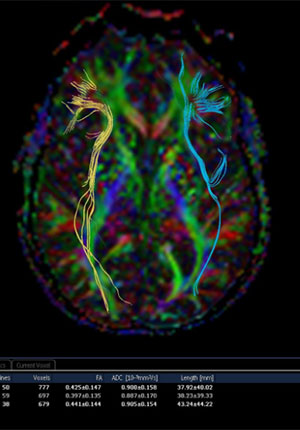
- Diffusion Tensor Imaging: In recent years, Diffusion Tensor Imaging (DTI) has emerged as a valuable tool for diagnosing TBI, particularly in cases involving subtle or diffuse injuries. DTI is a specialized form of MRI that maps the movement of water molecules in the brain’s white matter. This technique helps visualize and assess the integrity of neural pathways, making it especially useful for identifying injuries that disrupt brain connectivity. By detecting microstructural damage not visible on standard MRI scans, DTI provides critical insights into the functional impact of TBI and guides personalized treatment plans for recovery.
- Quantitative EEG examination (Qeeg): A qEEG (quantitative electroencephalogram) for a Traumatic Brain Injury (TBI) is a test that measures brain electrical activity using a specialized analysis to identify abnormalities in brain wave patterns, potentially indicating the presence and severity of a brain injury, particularly in cases where structural imaging like CT scans might not show significant damage; it can help assess the functional impact of a TBI by looking at brainwave frequencies, connections between different brain regions, and power distribution across different brain regions.
Main points about qEEG for TBI: By analyzing the electrical signals from the brain, a qEEG can detect abnormal changes in brainwave activity that may be associated with a TBI, even in mild cases where other diagnostic methods might not show abnormalities.
qEEG can be used to help diagnose a TBI, assess the severity of the injury, monitor treatment progress, and identify potential cognitive impairments associated with the injury.
Evoked Potentials: When assessing a traumatic brain injury (TBI), some commonly used evoked potentials are visual evoked potentials (VEP), brainstem auditory evoked potentials (BAEP), and somatosensory evoked potentials (SSEP), which measure the brain's response to visual, auditory, and tactile stimuli respectively, allowing clinicians to evaluate the integrity of the sensory pathways involved in each modality.
Why do we use evoked potentials?
- Detection of abnormalities:
Abnormalities in the latency (time taken for the response to occur) or amplitude (strength of the response) of these evoked potentials can indicate damage to the corresponding sensory pathways in the brain, which can be affected by a TBI. - Severity assessment:
The severity of the TBI can be partially assessed by the degree of abnormality seen in the evoked potentials. - Monitoring recovery:
Evoked potentials can be used to monitor a patient's recovery over time following a TBI, as improvements in the evoked potential waveforms may correlate with neurological improvement.
- Neuropsychological Testing: In addition to imaging, neuropsychological testing helps assess the cognitive impact of TBI. These tests measure a range of mental functions, including memory, attention, language, and problem-solving. This type of testing can reveal impairments in brain function that may not be visible on imaging tests. It is particularly valuable in diagnosing mild TBIs, where symptoms may not show up on traditional scans, but cognitive or emotional symptoms persist.
TBI is a multifaceted condition that can affect the brain in diverse ways. Different testing modalities—from imaging techniques like CT, MRI, qEEG, evoked potentials and DTI to neuropsychological evaluations—offer complementary perspectives on brain function and injury. Imaging can identify structural abnormalities, while neuropsychological tests assess cognitive and emotional impacts. By using a combination of these tools, we gain a comprehensive understanding of the injury, enabling more targeted and effective treatment strategies.
AUTHOR: Dr. Miguel A. Pappolla is a board-certified neurologist and pain medicine specialist in Houston, Texas, with expertise in traumatic brain injury and pain management. A full professor of Neurology at UTMB, he holds board certifications in five medical specialties and has authored over 100 highly cited research papers. With decades of experience as a consultant for the NIH and as a neuroscience educator, Dr. Pappolla continues to advance research and patient care.